In an attempt to identify Miocene stingray teeth recovered from the Pungo River (Miocene) and
Yorktown (Miocene-Pliocene) sediments at Aurora (Beaufort County, NC -- aka Lee Creek), extant species
are being studied. After cursory examinations of material available at the Smithsonian, it was
determined that more intense analysis of a greater number of specimens was required.
This is a small tropical ray from Florida and the Gulf of Mexico (known from Central America to the
Chesapeake). It inhabits shallow coastal waters. Snelson et al (1988) indicates that males mature at
20 cm DW (max 32.6 cm/1.6 kg) and females 24 cm DW (max 37.0 cm/2.2 kg). The population studied by
Peter Piermarini lives and breeds in Lake Jessup ( freshwater) of the St. Johns River in Florida.
Kajiura & Tricas (1996, citing Cook 1994) note that they feed upon small benthic crustraceans
(amphipods, mysids, isopods), polychaetes and invertebrates found in seagrass beds. During the
summer months they feed upon calcified brittlestar disks.
Peter Piermarini (pers com) noted that the Lake Jesup population appears to have no seasonal
shifts in diet. Stomach contents reveal a diet that is composed of viviparid gastropods, various
insect larvae (Chironomids and mosquito pupae) and small crustataceans (definitely no brittle stars).
Gastropod feet muscle and opercula are found in the gut, but the shell material is apparently spit out.
See Peter Piermarini page at
the Atlantic stingray
Dentition
The D. sabina dentition was traditionally viewed as having a crushing design in female specimens,
and clutching in males. Kajiura & Tricas (1996) demonstrated that the species in fact had a crushing
design, but in males, cuspidate teeth were present during mating season, giving them a seasonal,
grasping dentition.
The wear patterns on the teeth of specimen ELA95-10.5 suggest that the dentition may serve a grinding
function as well, at least on a seasonal basis. Terminology
 |
|
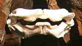
Fig. 1 ELA95-3.2 D. sabina
female dentition, 54 mm width |
As can be seen in figure 1, the occlusal surface of the upper tooth band is strongly depressed (concave) in
the mesial position (which corresponds with the teeth termed anterior) and raised (convex) in the lateral. The lower
tooth band matches this design by being raised in the mesial position and depressed in the lateral. The distal
regions of upper and lower are level.
For initial discussion purposes, each (upper and lower) tooth band will be divided into three regions - mesial,
lateral and distal. The mesial region will include 'symphyseal' and 'anterior' teeth, the lateral,' lateral' teeth and
the distal, 'posterior' teeth. It is unknown at this time if the teeth from various tooth band regions will display
sufficient odontological variations to be identified on an isolated tooth basis.
Prepared and studied specimens include:
| Specimen ID | Sex | Collected | Location | Disc
Width | Upr File
Formula | Lwr File
Formula |
| LJ-10/96 | Male | Oct. 96 | Lake Jesup | 22.8 cm. | 17-s-17 | 20-1:1-20 |
| LJ-3-31-97 | Male | Mar 31, 97 | Lake Jesup | 26.5 cm | 19-s-19 | damaged |
| ELA95-10.5 | Female | Jun 7, 95 | Lake Jesup | 37.0 cm | 20-s-20 | 20-1:1-21 |
| ELA95-3.2 | Female | Feb 16, 95 | Lake Jesup | 30.5 cm | | |
|
NOTE: The cleaning and curing process will cause the dentitions to assume a shape
that may not be totally natural or become distorted. The fleshy padding between the dental band and the jaws will
also be subject to shrinkage |
Specimen No. ELA95-10.5
This is the dentition of a large (37 cm DW) female captured June 7, 1995 in Lake Jesup. The dentition when dried
measured 73 mm in width. The upper tooth band is 37 mm in width and the lower, 34.5 mm.
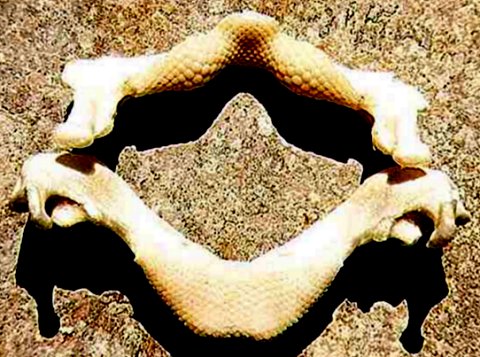 |
| Fig. 2 ELA95-10.5 D. sabina large female dentition, 73 mm width |
Upper Tooth Band. The dental formula is 20-s-20, forty-one teeth. There is a symphyseal,
the anterior teeth include file postions 1 - 6, the laterals 7 - 13 and the posteriors, 14 - 20.
The symphyseal & anterior teeth [s, 1-6] are small (1.2 x 0.9 mm,
meso-distal width x labio-lingual depth when noted) and less densely packed than the laterals.
There are 10 - 12 rows present which includes two which appear to be in the process of being shed,
and two sufficiently worn to be deemed 'functional'.
The lateral teeth [7 - 13] are the largest in the band reaching 2.0 x 1.8 mm (file 10). There are
10 - 11 rows, with 1.5 rows in a shed position and 3.5 in a functional.
Posterior teeth [14 - 20] have fewer rows (file 14 with 10 and 20 with 8) and gradually grow smaller
distally, 1.4 x 0.8 mm in position (file) 20. In file position 14, three rows are functional with another in a shed
position and in file 20, two are functional.
Lower Tooth Band. The dental formula is 20-1:1-21, forty-one teeth. The anterior teeth include
postions 1 - 6, the laterals 7 - 13 and the posteriors, 14 - 20/21. (The demarcation between laterals and
posteriors is currently rather subjective.)
The anterior teeth [1-6] are large (1.8 x 1.2 mm, in file 3) and densely packed. There are 15 - 16 rows
present which includes one or two which appear to be in the process of being shed, and four with
enough wear to be deemed 'functional'. These functional teeth are worn sufficiently to have a flat
occlusal surface and hexagonal shape -- giving teeth in these rows a myliobatoid-look (grinding
pavement teeth).
The lateral teeth [7 - 13] are small (1.2 x 1.0 mm in file 10). There are 11 - 14 rows, with one row
in a shed position and two in a functional.
Posterior teeth [14 - 20/21] have fewer rows (the most distal file has 8) and gradually grow smaller
distally, 1.0 x 0.7 mm in the last file. There appears to be three fuctional rows.
Specimen No. ELA95-3.2
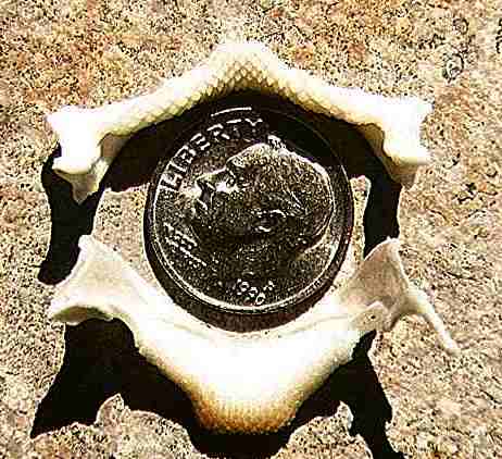
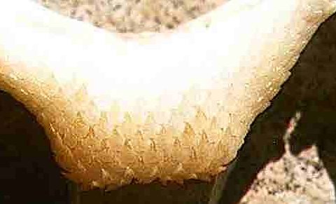
Specimen No. LJ-10/96 (Fig. 3)
This is the dentition of a (22.8 cm DW) male captured in October 1996 in Lake Jesup.
The dentition when dried measured 33 mm in width. The mandibular cartilage was damaged during
removal resulting in the loss of about 2 mm from the right side. The upper tooth band is 20 mm in
width and the lower, 18.5 mm. This dentition reflects the seasonal dimorphism described by
Kajiura & Tricas (1996). Within each file, there is an abrupt change in tooth morphology
— the teeth become significantly more cuspidate lingually.
 |
|
| Fig. 3 LJ-10/96 D. sabina male 22.8 cm width |
Upper Tooth Band (Fig. 4). The dental formula is 17-s-17, thirty-five teeth. There is a symphyseal,
the anterior teeth include file postions 1 - 7, the laterals 8 - 14 and the posteriors, 15 - 17.
The symphyseal & anterior teeth [s, 1-7] are high-cusped and small (c1.0 mm in width).
There are seven rows present which appear to include three which are functional. In row five, the
cusps become clearly larger.
The lateral teeth [8 - 14] are the largest in the band (file 10) reaching c1.3 mm in width and
low-cusped. There are seven rows, with 2.5 appearing to be functional. Begining in row four,
the teeth have higher cusps.
Posterior teeth [15 - 17] are small and non-cuspidate. Arranged in five rows, they gradually
grow smaller distally (c0.8 mm in file 17). Two rows show evidence of being functional and a
uspidate morphology appears in row three.
NOTE: The file breakpoints between tooth-types is an approximation only.
The teeth are small and were observed in situ. The breakpoints were based on observable tooth structure
and tooth band location. These may be revised in the future, but it is unlikely that they will vary by more
than one file position. For the same reason, tooth measurements could not be reliably made and can
be off by 0.2 mm. .
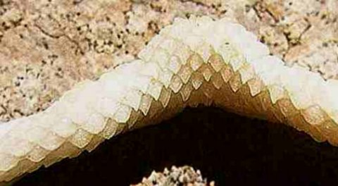 |
Fig. 4 LJ-10/96 D. sabina male, upper tooth band
Illustrated is the symphyseal, right hand files 1 - 16 and left hand files 1 - 12.
Anterior to top. |
Lower Tooth Band (Fig. 5). The dental formula is 20-1:1-20 -- forty teeth. The anterior
teeth include positions 1 - 7, the laterals 8 - 16 and the posteriors, 17 - 20.
The anterior teeth [1-7] are high-cusped and the largest in the tooth band (width c1.0 in file 1
& c1.2 in file 4). There are 9 teeth in file 1 decreasing to 8 in file 7. One row is being
shed, 2.5 appear functional and they become higher cusped in row 5.
The lateral teeth [8 - 16] are smaller c1.0 mm in width) and are currently deemed non-cuspidate.
There are seven rows (file 12), with three functional (may include teeth in a shed position).
Teeth become cuspidate in row four.
Posterior teeth [17 - 20] have fewer rows (the most distal file has 6) and gradually grow smaller
distally (c0.7 mm in the last file). There appears to be three functional rows and teeth become cuspidate
in row three or four.
 |
| Fig. 5 LJ-10/96 D. sabina male, lower tooth band |
Specimen No.LJ-3-31-97
This is the dentition of a (26.5 cm DW) Lake Jesup male captured in March 1997. The right hand side of this dentition
was severed during removal, but would have been of slightly wider than LJ-10/96 (c36 mm in width). The
upper tooth band is 22 mm in width, but the lower cannot be determined. In this dentition, all rows contain
high-cusped teeth. Within each row, there is an abrupt change in tooth morphology -- the teeth become
significantly more cuspidate. Multiple rows are missing from the labial margin of the tooth bands
(particularly the upper), this may possibly be the result of pectoral biting during mating.
Upper Tooth Band. The dental formula is 19-s-19, thirty-nine teeth (files). There is a symphyseal,
the anterior teeth (subjectively) include file postions 1 -7, the laterals 8 - 12 and the posteriors,
13 - 19. The symphyseal & anterior teeth [s, 1-7] are high-cusped and small (c1.0 mm in width).
The lateral teeth [8 - 12] are the largest in the band reaching 1.3 mm in width. Posterior
teeth [15 - 17] are small and fully cuspidate.
Lower Tooth Band. Neither side of the tooth-band is complete -- row and file counts
cannot be provided. It would be highly subjective to differentiate anterior teeth from laterals, however
in files RH-14/LH-11, the tooth cusps become markedly lower, suggesting the begining of the posterior
tooth-file groups.
A Fossil Record?
The D. sabina tooth design has not been noted in the Lee Creek Fauna (Miocene,
North Carolina). However, odontologically-similar teeth are represented in the Sharktooth Hill
Fauna (Miocene, California). Considering the current distribution of this species, and in the absence
of connected Americas, there is no reason to fully discount this possibility.
References
Bigelow, H. B. & Schroeder, W., C., 1953. Fishes of the Western North Atlantic.
Sawfishes, Guitarfishes. Skates and Rays. Memoires of the Sears Foundation for Marine
Research, no. 1, part 2.
Kajiura, Stephan M. & Tricas, Timothy C., 1996. Seasonal dynamics of dental
dimorphism in the Atlantic Stingray Dasyatis sabina. Journal of Experimental Biology,
pp 2297-2306.
Snelson, F.F., Williams-Hooper, S.E. and Schmid, T.H., 1988. Reproduction and ecology of
the Atlantic stingray, Dasyatis sabina, in Florida coastal lagoons. Copeia. pp 729 - 739