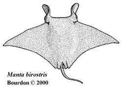
In an attempt to identify fossil Manta-like teeth from Lee Creek (Miocene
deposits from North Carolina), an attempt was made to compare them with those from an
extant Manta ray. After a lengthy search, a tooth band was finally obtained. The below
data provides a summary of a preliminary study of that dentition.
The Dentition
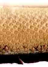
Ostensibly homodont, the dentition of the manta is made up of many alternating files of small, peg-like teeth.
The studied example, although incomplete, had 196 files. Except for distal files, each contained
5 or 6 developed teeth. (An accompanying chart details these file and row counts.) In addition,
each file showed evidence of the formation of one or two new teeth. The crowns are widely spaced,
but the bulbous roots causes the teeth to be tightly juxtaposed. The dentition as received measures
207 x 15 mm.
Due to the homogeneous nature of the manta tooth band, functional position terms, such as anterior, lateral and
posterior, have little use when discussing particular teeth. Under normal circumstance, it would
be appropriate to refer to teeth based upon their relative position to the symphysis. However,
when the studied tooth band was removed from the lower jaw, the right hand side was incomplete.
Although an assumption has been made as to the position of the symphysis based upon
attached skin, the actual tooth positions cannot be positively established. Therefore, positional
references will be made by indicating the inferred position relative to the symphysis (i.e., LH-34)
and the absolute one-up position (i.e., 80) from the left-hand corner of the mouth. To see itemized
details regarding tooth counts and mesurements, see:
Dentition Details.
Note: All dentition images are oriented similarly. Labial edge
towards the bottom and lingual towards the top. This causes the right side to be on the left
and vice versa. Since the absolute tooth positions are one-up from the distal left hand tooth
(right in image), they will be listed similarly (i.e., 127-91). The position relative to the
symphysis will be user friendly. presented as visible in the particular image. Therefore,
looking at a group of right-hand teeth (left side in image), the positional reference would
be RH 45-33. If these teeth were on the left, this notation would be LH 33-45.
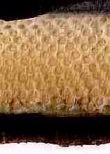
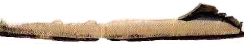 |
| Fig. 1 M. birostris (lower) tooth band, width 207 mm |
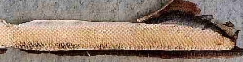 |
Fig. 2. Tooth band enlargement.
Includes all of the left-hand files (113).
(Labial margin at bottom) |
 |
 |
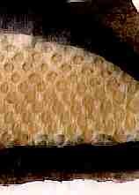 |
Fig. 3
Files LH-32 - 45,
pos 82 - 69 |
Fig. 4
Files LH-70 - 84,
pos 44 - 30 |
Fig. 5
Files LH-97 - 111,
pos 17 - 3 |
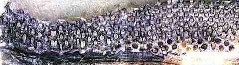 |
Fig. 6
Files RH-83 - 65,
pos 196-178, [blue dye] |
Fig. 7
Files RH-64 - 43,
pos 177-156 [blue dye] |
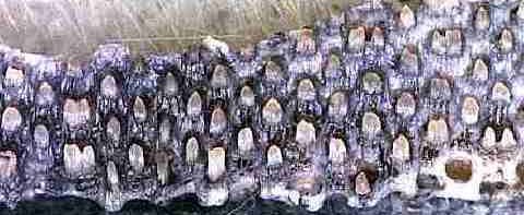 |
Fig. 8 Files RH-78 - 62, pos 191-175 [blue dye]
(Note laterally expanded crowns in files 76 & 64.) |
Examined Teeth
Eight teeth were removed from shed positions of the dentition for closer study. They revealed
a surprising degree of variability in both shape and size. A cursory examination found no
relationship between tooth shape and file position. Additional study would be required to
determine if there are positional differences reflected by these teeth or if a particular germ
produces these variations. The below examples should prove useful in illustrating the
potential variability of these teeth.
Without a reliable means of differentiating teeth by position, and unable to determine if sexual
dimorphism is relevant, the teeth examined so far are being termed "T" (typical), "D" (denticle-like) and
"S" (skewed) -Type.
Typical Teeth - Type - "T"
The majority of teeth in this dentition were typical of illustrations seen in other sources. During this
preliminary study, teeth ascribing to this basic design are being referred to as Type "T".
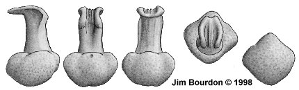 |
Fig 9. T-type (181/RH-68)
height 1.7, width 1.1 and depth 1.1 mm |
T-type (181/RH-68 - Figure 9). This specimen represents the typical manta tooth.
The root is bulbous, as wide as deep and weakly flattened apically. A single foramen is
situated high on the labial protuberance. Viewed basally, the profile is relatively square
and the lateral segments blend evenly with the labial and lingual protuberances. Much
narrower, and wider than deep, the crown stem rises vertically from the root. The cusp extends
lingually from the stem reaching a position nearly perpendicular to the lingual margin of the
root. A trough is created between the transverse margins of the cusp and extends down the
labial face of the stem, narrowing basally. Labio-lingual ridges are present in the cuspal
area of the trough. [Specimen measures: height 1.7, width 1.1 and depth 1.1 mm. Original
illustration 68-D, 20X]
T-type (167/RH-54). This tooth similar to 181/RH-68. [Specimen measures:
height 1.4, width 1.0 and depth 1.0 mm]
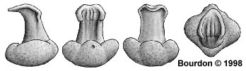 |
Fig. 10. T-type (80/LH-34).
height 1.3, width 1.2 and depth 1.1 mm |
T-type (80/LH-34 -- Figure 10) Bulbous but apically flattened root, approximately as wide
as deep. Labial and lingual protuberances of the root are evident, the labial one contains
a single foramen. Viewed apically or basally, the labial and lingual protuberances create a relatively
square appearance. Much narrower than the root, the crown stem is set into the occlusal face
of the root. The stem is broader than deep. A distinct trough runs down the labial face of the stem,
narrowing basally. The lingual face of the stem has weak apico-basal ridges. Apically, the
crown stem bears a lingually directed enameloid cusp which extends back, nearly to a
perpendicular line from the lingual margin of the root.. The occlusal and labial face of the cusp
has several lingually directed enameloid ridges. These ridges appear to bear granular
ornamentation, particularly in the area where the cusp joins the crown stem. Viewed occlusally,
the cusp is slightly broader then the stem and is leaf-shaped in appearance. [Specimen
measures: height 1.3, width 1.2 and depth 1.1 mm. Original illustration 68-A, 20X]
T-type (41/LH-73) This tooth very similar to 80/LH-34 except foramen not observed.
[Specimen measures: height 1.3, width 1.0 and depth 1.0 mm]
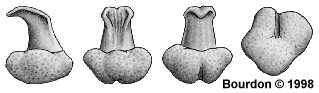 |
Fig. 11. T-type (67/LH-47)
height 1.2, width 1.0 and depth 1.1 mm |
T-type (67/LH-47: Figure 11) Bulbous root with distinct labial protuberance and no
visible foramen. Unique to this specimen is a relatively strong nutrient groove in the lingual
hemisphere of the root. The crown stem is erect, bears a lingually directed cusp and is weakly
inset into the apical face of the root. The enameloid cusp, leaf-shaped when viewed occlusally
bears, labio-lingual ridges which extend down the labial face of the stem. [Specimen measures:
height 1.2, width 1.0 and depth 1.1 mm. Original illustration 68-C, 20X]
Denticle-like Teeth - Type - "D"
So far, this tooth-type is represented by a single specimen. Had it not been removed from the
dentition, it would have been thought to be a dermal denticle with an ornamented cusp. The gradual
lingual curvature of the stem and cusp has not been reflected in other studied teeth from this dentition.
This is likely to be the result of very short stem combine with a labio-lingually elongated cusp.
For its denticle-like design, it will be referred to as Type "D".
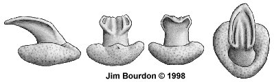 |
Fig. 12. D-type (22/LH-92)
height 1.1, width 1.2 and depth 1.6 mm |
D-type (22/LH-92: Figure 12) Broad, apically flattened root with little observable
characteristics; the basal profile is tear-dropped. A labial foramen was noted but very weak,
and the overall appearance is that of a dermal denticle base. The crown stem is short, labially
directed and weakly inset into the occlusal face of the root. The crown’s cusp further adds to the
denticle-like design, extending well beyond the lingual margin of the root. The occlusal face of
the cusp is concave, and although worn, enameloid ridges are present. [Specimen measures:
height 1.1, width 1.2 and depth 1.6 mm. Original illustration 68-B, 20X]
Skewed Root Teeth - Type - "S"
The initial reaction when viewing this tooth morphology (ref Fig. 13) is that it reflects a multi-crowned design.
However, in the specimen from tooth position 194/RH-81, there appears to be a single stem/crown. Until
additional laterally elongated teeth can be studied, they will be categorized by their skewed root and
be referred to as Type "S".
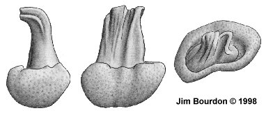 |
Fig. 13 S-type (189/RH-76)
height 2.2, width 1.8 and depth 1.4 mm |
S-type (189/RH-76: Figure 13) This illustration represents a laterally expanded design which
appears to include a second stem. This tooth is not currently thought to be pathological. Viewed
laterally, the root appears bulbous. Labially, a protuberance is not evident; rather, there appears
to be evidence of a third, laterally compressed lobe. No foramina were noted. Lingually, a laterally
expanded protuberance appears to be present. The crown is made up of two, joined stems. The
stems are erect with lingually directed cusps and are inset into the apical face of the root. The cusps
bear, ornamented, labio-lingual ridges which extend down the labial face of the cusp. [Specimen
measures: height 2.2, width 1.8 and depth 1.4 mm. Original illustration 68-E, 20X]
S-type (194/RH-81) This tooth is similar to 189/RH-76 but is single-cusped. The crown
and root appear diagonally skewed, making it difficult to orient it’s actual position. [Depending
how the tooth is oriented, the specimen measures: height 2.0, width 0.8 and depth
1.5 or 2.0 x 1.3 x 1.3 mm
The Manta Tooth
The manta crown design, although unusual, is not unique. Simple, peg-like teeth are also
employed by Mobula japanica (MULLER & HENLE, 1841). Notarbartolo
di Sciara (1987) depicts the latter’s teeth. [Although M. japanica teeth are peg-like,
the roots are not bulbous (being either laterally or labio-lingually compressed) and have
a fully formed nutrient groove which runs the (labio-lingual) depth of the root. The crown’s
cusp reflect weak to strong lateral cusplets and the root has neither a labial nor lingual protuberance.]
Root. The root is very broad, both labio-lingually and mesio-distally, extending well beyond
the crown stem. It is usually high, giving a bulbous appearance, but can be apico-basally
compressed, giving a denticle-type appearance. The lateral lobes of the root merge with
labial and lingual protuberances to give the root its bulbous appearance. Usually these root
segments are weakly differentiated. In figure 11, a short nutrient groove is clearly visible between the
lateral lobes. Figure 13 shows polyaulacorhizous root characteristics, providing evidence of a
myliobatoid origin of the odontological design. In many teeth, a weak foramen may
be observed on the upper face of the labial protuberance.
Crown Stem. Characteristic of these teeth is the high stem which positions the cusp well
above the root. The stem is usually oriented perpendicular to the root, but may be directed
lingually (figure 12). The stem is usually inset to some extent into the apical face of the root
and has a concave groove running down the labial face which may bear extensions of the
cusp’s labio-lingual ridges. The stem is usually (laterally) wider than deep and often appears
'pinched' when viewed labially.
Cusp. The enameloid covered cusp extends lingually from the stem and appears
leaf-shaped from an occlusal aspect. Viewed occlusally, there is a trough between the wide
ridge of the transverse margin which extends down the root stem, narrowing basally. This trough
bears multiple labio-lingual ridges that may continue down the face of the stem.
Cusp Wear The occlusal surface of the crown shows wear (albeit weak), without an
opposing dentition (upper) -- this struck me as surprising. Without a known shed rate, the relative
amount of wear can not be determined. I asked Rachel Alexander if the contact surface of the
tooth band had denticles, but she said it was "smooth, with no evidence of teeth or denticulate
processes." She tendered two possiblities: the teeth may be used in an attempt to remove
parasites from the upper jaw, or possibly, on other individuals during courtship.
References
Bigelow, H.B. and W.C. Schroeder 1953. Part 2. Sawfishes, Guitarfishes, Skates
and Rays; Chimaeroids In: Fishes of the western North Atlantic.. Memoires of the
Sears Foundation for Marine Research, Yale University, New Haven
Cappetta, H.1987. Chondrichthyes II: Mesozoic and Cenozoic Elasmobranchii.
Handbook of Paleoichthyology. Gustav Fischer Verlag, Stuttgart and New York