In January 1997, an informal project was spawned, quite by accident (heard that
before?). The seeds were sown so-to-speak during e-mail conversations between Henry Mollet,
volunteer at the Monterey Bay Aquarium (MBA) and Jim Bourdon (currently researching Miocene batoids).
The original question was:
"Is D. violacea represented in the Lee Creek fauna?" Unable to obtain a dentition for
comparative purposes, Scotty Greenwald, an aquarist at the Monterey Bay Aquarium reclaimed
shed teeth teeth from the bottom of a tank with pelagic stingrays.
 Pteroplatytrygon violacea
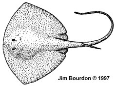
This is a small (to 80 cm/32 inches disc width) pelagic ray of worldwide distribution
in tropical and temperate waters. It feeds on squid, herring, mackerel, octopus, and
crustaceans. The disc is trapezoidal in shape, purple in color and bears thorns on the back and tail.
A Studied Dentition
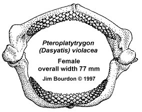
The illustrated dentition is from a 47 inch (total length) ray caught by fishermen in deep waters off
Santa Rosa Island, California on October 5th, 1996. Although not positively identified at the time
of capture, the ray was described as having a dark purple back with some light spots and a light
purple belly. They went on to note that it had rounded tips, a recurved back, a long tail and a
Bat ray-looking head. The overall description, time and location of capture, long thin stinging
barb (also retained) and the teeth themselves leave little doubt that it was a pelagic stingray.
 NOTE: The discussion of this particular dentition is based on teeth in-situ,
it was not torn apart to properly measure and study the teeth. Various teeth in the process of being
shed were removed and measured, but most measurements could only be made directly from the dentition
using a millimeter-scaled ruler and a loupe. Although the authors feels these measurements are useable,
they may not be as accurate as implied by their notation in tenths of a millimeter.
NOTE: The discussion of this particular dentition is based on teeth in-situ,
it was not torn apart to properly measure and study the teeth. Various teeth in the process of being
shed were removed and measured, but most measurements could only be made directly from the dentition
using a millimeter-scaled ruler and a loupe. Although the authors feels these measurements are useable,
they may not be as accurate as implied by their notation in tenths of a millimeter.
The Jaws
In this monognathic heterodont dentition, all teeth could be said to be cuspidate. The higher-cuspsed
teeth are in the medial position, growing lower distally. Although upper and lower teeth are very similar,
they appear to be distinguishable in lateral profile.
As illustrated, the jaws are 77 mm wide and 58 mm high, the internal width (narrowest) is 48 mm.
The upper dental band is 51 mm wide and the lower, 44 mm. The teeth in this dentition compare
favorably with a female specimen at the Monterey Bay Aquarium.
Tooth design
The tooth bands from this species are unlike those of most stingrays. Not only do both sexes
have cuspidate teeth, but there are no crushing teeth in the lateral files.
The medial teeth have a traditional dasyatid (male) cuspidate (grasping) crown which is
derived from an elongated cusp, rising from the lower crown and directed lingually. Viewed labially,
these teeth have a distinctly pointed crown.
The lateral teeth, however, do not have the globular crown of a crushing dentition as is
present in most stingrays. The labio-lingually compressed upper regions of the crown has produced
a high, usually (meso-distally) elongated, transverse crest -- a cutting dentition. The crest is enhanced
by enameloid ridges which would produce a serrate-like cutting edge. Certain anterior lateral teeth
may be cuspidate when viewed labially.
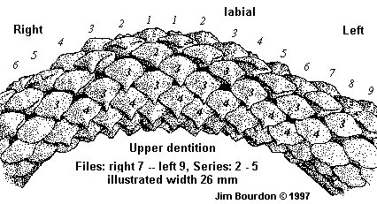
Tooth Files
Dental Count Upper 17-1-17 (34 files); Lower 19-S-19 (39 files).
 |
| Pteroplatytrygon (Dasyatis) violacea |
Cusps. For upper teeth, files #1 - 3/4 are high-cusped, files #4/5 - 7 low-cusped. In the lower teeth,
files #S - 1 are high-cusped, 2-7 are low-cusped. The remaining files for each dental band, have a high
transverse ridge which begin with a cusp-like apical angle in position #8 and gradually decrease in
height distally.
File Groups When viewing the teeth as a dentition, the files appear to be segregated into two groups,
medial and lateral. (Please note that there will be a further refinement
when discussing the teeth themselves.)
Medial teeth [files S(1) - 7]. In this dentition, these are the cusp bearing crowns and are
of similar size (2.9 - 3.0 mm in width and about 2 mm in depth) although the symphyseal and file #1 are
slightly narrower (laterally compressed).
Lateral teeth. In the upper dental band, U8 - 11 are the broadest teeth of the dentition,
measuring to 3.3 mm in width. Beginning with U12 (2.9 mm) the teeth diminish in width to U17 (2.0 mm).
In the lower band, L8 - L9 are 2.6 - 2.8 mm in width, U10 (2.4 mm) and continuing to narrow to U19 (1.8/2.0 mm).
Although few teeth could be measured properly, several removed teeth show a significant labio-lingual
compression of the lateral teeth distally. Selected width x depth measurements include:
UL5: 3.0 x 1.8, UR10: 3.3 x 1.9, UR15: 2.4 x 1.5 and LL5: 3.0 x 2.1, LR10: 2.4 x 1.8 and LR15: 2.2 x 1.3 mm.
Tooth Series
The series count includes a single seed or germ tooth and a to-be-shed tooth.
Upper file #1 = 9, file #9 = 8, file #17 = 7 teeth and Lower file #1 = 9, file #9 = 9, file # 19 = 7 teeth.
Functional Teeth. In this dentition, the "first" (labial most) upper series is in the process
of being shed, and the second appears functional. In the lower teeth, the second series appears
worn and the third appears to be the primary functional series.
Tooth Wear: The series being shed showed no evidence of the cusps which were
visible on the functional and replacement teeth. [A fossil tooth would likely not reveal the
cuspidate design of these teeth.]
Bicuspidate Several teeth show evidence of a bicuspidate condition. In this dentition
they were relegated to files 4 - 8. This condition was noted in replacement series (as high has series 7)
suggesting this is not a wear condition but an odontological characteristic of the species.
Tooth Characteristics
There appear to be several morphological constants in these teeth - many are
characteristic to the family, others are less common.
- All non-pathological teeth have bilobate, lingually directed roots with a single central foramen.
The teeth have not been stained or coated to enhance details, but lateral foramina, on the interior
surface of the lobes, have not been observed. A second foramen in the nutrient groove has not been
noted although at least one central pore appeared to be made up of two adjoining foramina.
- The lateral profile of most cuspidate teeth are of typical dasyatid design. Some cuspidate
teeth have an unusual feature: they are cuspidate but the slope of the lingual face is labially
oriented -- a cuspidate transverse crest.
- The labial face of all teeth have a consistent characteristic, there are narrow, closely-spaced,
intersecting enameloid ridges running from the labial visor to the apex of the cusp. This creates
a pitted appearance, which becomes slightly more elongated apically.
- In most teeth, there is a transverse depression on the labial face. In cuspidate teeth, it
is often weak and located between the visor and cusp portions of the crown. In posterior and some
lateral teeth, it is often very deep, helping to create the prominent transverse crest. Most teeth
reflect longitudinal grooves on the upper lingual face in the area of the transverse ridge.
- These teeth often have a notched lingual rim which gives the appearance of a two uvula, one
above each root lobe. This is likely a result of the dense packing of these teeth.
General Description
Male and female pelagic stingray teeth consist of a bilobate root and a cuspidate crown. The
teeth are small, ranging in width (mesio-distal) from 2 to 4 mm and depth (labio-lingual) from
1 to 2-1/2 mm. A moderately deep and wide nutrient groove is present between the two, lingually
directed, lobes. There is a single central pore which usually contains a single foramen. Lateral
foramina, on the interior surface of the lobes, have not been observed. The root lobes do not
extend beyond the lateral margins of the crown.
Anterior and lateral teeth of both sexes have cuspidate crowns, in contrast to the typical globular
design associated with most stingrays. The medial teeth have strong, lingually directed cusps,
which are similar to the "typical" high-cusped male stingray tooth -- such as those from
Dasyatis americana HILDEBRAND & SCHROEDER, 1928,
D. sayi (LESUER, 1817) and
D. sabina (LESUER, 1824).
Unlike the cited examples, the labial face does not have strong enameloid ridges or a
medial depression running the length of the cusp. In lateral teeth, the cusp is weaker and
more upright. In lieu of a cusp, the posterior teeth have a prominent, laterally elongated,
transverse crest which adjoins a strong transverse depression on the labial face. On the
lingual face, the transverse crest has longitudinal ridges. Medial and lateral teeth have a
weak depression on the labial face which is located at the juncture of the crown base and the cusp.
The labial face of all teeth have a pitted appearance which results from the depressions created
by numerous small meandering enameloid ridges. No teeth have a "cutting edge" as in many
shark or skate teeth. Rather, the acute angle of the transverse edge of the cusp and transverse
crest have a serrate edge created by the numerous enameloid ridges.
As a dasyatid, there is a unique feature represented in these teeth -- the development of a
second cusp. An incipient reflection of this characteristic can be seen in many teeth, usually
as a longitudinal depression in the lingual face of the crown. Eight percent of the teeth, originating
in medial and lateral positions, have a second cusp. These bicusped teeth appear sporadically
within the dentition, and are present in a file of teeth which are otherwise single-cusped. Unlike the
mobulids, the other family developing additional cusps, this bicuspidate condition in the pelagic
stingray is not associated with the mesio-distal elongation of the crown.
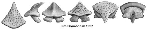
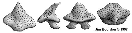
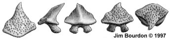
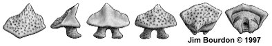
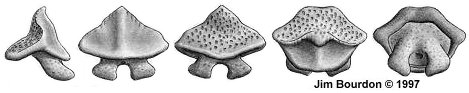
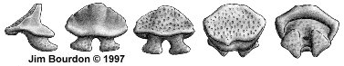
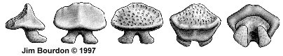
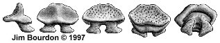
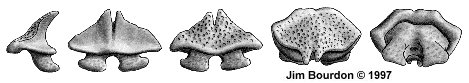
| NOTE: All teeth were originally illustrated at 10X
and have maintained relatively similar enlargement factor (to one another) when
digitized. From left to right, the aspects are: lateral, lingual, labial, occlusal and
basal. In certain cases, a cuspal view has been added to the left or one or more
aspects may have been omitted. |
Bi-cuspidate Teeth
An unexpected morphological condition in this study was the presence of a second cusp in the teeth
of this species.
 |
| Bi-cusped Tooth hgt = 2.1, wid = 3.2, dep = 2.0 mm |
References
Cappetta, H, 1970. Les sélaciens du Miocène de la région de Montpellier.
Palæovertebrata (1970), mém, ext.: pp 1-139.
Cappetta, H. 1987. Chondrichthyes II. Mesozoic and Cenozoic Elasmobranchii
Compagno, L. & Heemstra, P. 1984. Himantura draco, A new species of
stingray from South Africa, with a key to the DASYATIDAE and
the first record of Dasyatis kuhlii from Southern Africa.
Deynat, P & Séret, B 1996 Le revêtement cutané des raies, I - Morphologie et
arrangement des denticules cutanés.
Herman, J., Hovestadt-Euler, M., Hovestadt, D.C. & Stehmann, M. 1994 and 1995.
Contributions to the Study and Comparative Morphology of Teeth and Other Relevant
Ichthyodorulites in Living Supra-specific Taxa of Chondrichthyan Fishes, Part B:
Batomorphii No. 1a and 1b Order Rajiformes - Suborder Rajoidei - Family:Rajidae.
Bulletin de L'Institut Royal des Sciences Naturelles de Belgique. Volume 64, pp 165-207
and volume 65, pp 237-307.
Kajiura, S. & Tricas, T. 1996. Seasonal dynamics of dental sexual dimorphism in
the Atlantic stingray Dasyatis sabina. The Journal of Experimental Biology.
pp 2297-2306.
Nishida, K. 1990. Phylogeny of the suborder Myliobatidoidei. Memoirs of the Faculty of
Fisheries, Hokkaido University 37 (1/2): 1-108.
Nishida, K. & Nakaya, K. 1990. Taxonomy of the genus Dasyatis
(Elasmobranchii, Dasyatidae) from the North Pacific pp 327-346. Elasmobranchs as Living Resorces:
Advances in the Biology, Ecology, Systematics, and Status of the Fisheries,
H. L. Pratt, S. H. Gruber, T. Taniuchi (eds.). NOAA Technical Report NMFS 90. U.S. Department of Commerce.
Radinsky, L. 1961, Tooth Histology as a Taxonomic Criterion for Cartilaginous Fishes.
____, pp 73-92.
Stehmann, M. , 1981. FAO Species Identification Sheets for Fisheries Purposes,
Eastern Central Atlantic, Batoid Fishes.
Welton, B & Farrish, R. 1993. The Collector's Guide to Fossil Sharks and Rays
from the Cretaceous of Texas
GOTO: Mollet home page
For additional details on this research, to tender suggestions, or whatever, contact one of us:
Henry Mollet,
Jim Bourdon or
Scott Greenwald