The shark tooth fossil record throws many curve balls at the collector,
one of which is the possibility that a specimen reflects a 'pathological'
condition. I have neither the time nor inclination to research the
proper utilization of the term 'pathological' (if there is one) in this
context. Therefore, I will employ this term to mean that a tooth (from
a given or assumed file position) is of non-standard design, probably as
a result of damage to the 'tooth germ' that produces teeth in a given
file.
File Splitting versus Injury
 Over the years, certain natural 'abnormalities' have been deemed pathologies. The best
example might be Gudger (1933) who attributed certain tooth variations in rays to
pathologies, when they were merely the natural manifestation of file splitting. I can
not insure the reader that similar oversights might not occur on this page, but every
attempt will be made to avoid this trap.
Over the years, certain natural 'abnormalities' have been deemed pathologies. The best
example might be Gudger (1933) who attributed certain tooth variations in rays to
pathologies, when they were merely the natural manifestation of file splitting. I can
not insure the reader that similar oversights might not occur on this page, but every
attempt will be made to avoid this trap.
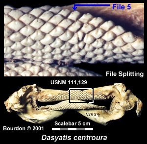
In the accompanying image of the Roughtail Stingray, the fifth upper left file
is in the process of splitting. The oldest tooth (at the labial margin) has a
normal design. The following tooth is laterally expanded and by the third row, a
second cusp is clearly visible. It is possible that these teeth still share a
common root. In row five, there are now two teeth occupying the old (now expanded)
file 5 position.
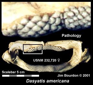 Unlike 'file splitting', tooth germs may be injured thus creating abnormal teeth.
These pathologies are likely to continue over time, showing their presence
in the tooth-band. The accompanying image of a Southern Stingray reflects germ
damage which could be associated with a fish hook injury. Despite the obvious
deformities of these teeth, it could be difficult to distinguish these teeth if
represented by a isolated fossil tooth.
Unlike 'file splitting', tooth germs may be injured thus creating abnormal teeth.
These pathologies are likely to continue over time, showing their presence
in the tooth-band. The accompanying image of a Southern Stingray reflects germ
damage which could be associated with a fish hook injury. Despite the obvious
deformities of these teeth, it could be difficult to distinguish these teeth if
represented by a isolated fossil tooth.
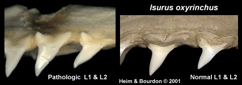
The below images shows the result of tooth bud damage in the Shortfin Mako.
In the two pathologic files (1st and 2nd upper lateral),
all the replacement teeth show the same distortions .
(Note the damage to the jaw cartilage above the teeth.)
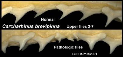
A comparison between a normal Spinner shark dentition and one with a serious
pathological condition.
Other Abnormalities
There are undoubtedly various other conditions, which can result in pathological
teeth, however no attempt will be made expand upon this point. It should be noted
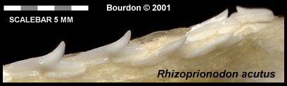
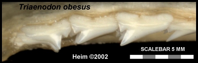
that a tooth band may reflect an abnormality not observable in a fossil tooth. The
accompanying images of Milk and Whitetip Reef Shark dentitions reveals that the
posterior tooth is misdirected. The Bourdon collection has a Galeorhinus with
two reversed files (adjoining) and Gordon Hubbell advised that he's seen this
phenomenon a couple times. In one case, it was the fourth or fifth file of a Tiger
Shark that had reversed teeth.
The below dentition of a Grey Reef shark appears to be missing file 6, at least
there is an abnormal diastema in the upper laterals. This space has resulted in the
teeth of the adjoining file to have abnormally elongated distal shoulders.

Abnormalities versus Variations
 It is well known that teeth from a particular file position can vary to some degree
based on sex, age, region and between individuals. The symphyseal region seems
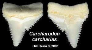
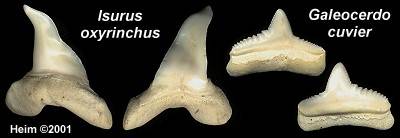
particularly prone to producing multiple 'morphologies'. These images provide
examples of pathological great white, mako and tiger shark teeth.
It is well known that teeth from a particular file position can vary to some degree
based on sex, age, region and between individuals. The symphyseal region seems
particularly prone to producing multiple 'morphologies'. These images provide
examples of pathological great white, mako and tiger shark teeth.
In the case of teeth that resemble the symphyseal designs, seeing the actual
dentition might be required to determine if certain specimens are
the result of a pathology or merely an unusual variation of a file position.
Fossil Specimens

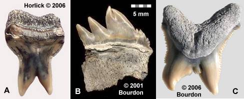 |
A - Physogaleus contortus, Lee Creek, Alex Horlick collection
B - Notorynchus cepedianus, Lee Creek
C - Carcharhinus sp, Lee Creek, John Timmerman collection
|
References
Gudger, E. W. 1933 Abnormal Dentition in Rays, Batoidei
Journal of the Elisha Mitchell Scientific Society. Vol 49(1), pp 57-96, 1 plate
|