EXTANT BATOIDS
Pelagic Stingray Shed Teeth
Jim Bourdon, Scott Greenwald and Henry Mollet
Illustrated by Jim Bourdon, Copyright © 1997
Objectives
From the outset, many objectives were clear, others emerged has the study progressed.
They currently include:
- Determine if characteristics exist to distinguish male from female,
upper from lower and anterior from lateral and posterior.
- Establish tooth shed rates for the species
- Determine if changes in odontology occur during breeding season.
- Establish the validity of the bi-cuspidate morphology of many of these teeth
Tooth Characteristics
There appear to be several morphological constants in these teeth - many are
characteristic to the family, others are less common.
- All non-pathological teeth have bilobate, lingually directed roots with a single central foramen.
The teeth have not been stained or coated to enhance details, but lateral foramina, on the interior
surface of the lobes, have not been observed. A second foramen in the nutrient groove has not been
noted although at least one central pore appeared to be made up of two adjoining foramina.
- The lateral profile of most cuspidate teeth are of typical dasyatid design. Some cuspidate
teeth have an unusual feature: they are cuspidate but the slope of the lingual face is labially
oriented -- a cuspidate transverse crest.
- The labial face of all teeth have a consistent characteristic, there are narrow, closely-spaced,
intersecting enameloid ridges running from the labial visor to the apex of the cusp. This creates
a pitted appearance, which becomes slightly more elongated apically.
- In most teeth, there is a transverse depression on the labial face. In cuspidate teeth, it
is often weak and located between the visor and cusp portions of the crown. In posterior and some
lateral teeth, it is often very deep, helping to create the prominent transverse crest. Most teeth
reflect longitudinal grooves on the upper lingual face in the area of the transverse ridge.
- These teeth often have a notched lingual rim which gives the appearance of a two uvula, one
above each root lobe. This is likely a result of the dense packing of these teeth.
File Groups
When studying the dentition only, there appeared to be two general tooth-types, medial and lateral.
In studying shed teeth however, there appears valid arguments for assigning three groups: anterior,
lateral and posterior. Anterior teeth have elongated cusps, laterals appear cuspidate when viewed
labially only, and posteriors have a high, level, laterally elongated and labio-lingually compressed
transverse crest.
Specimen Numbering
At the time of this writing, these specimens have not been assigned to a particular collection and
are being tracked only by collection date. The preliminary numbering system is
YR-ORG-SP-MMDD-NN (i.e., 97-MBA-DV-0117-22) and has a shorthand notation of MMDD-NN.
An arbitrary "tooth-type" designation has not been developed. For discussion purposes, the
nomenclature employed only refers to the illustration's number. This should be a temporary
condition. Described tooth-types which are not illustrated are assigned a one-up numbering system
with a "NYI" (not yet illustrated) prefix.
Collected Lots
This study began with a sample of 121 teeth that represented three weeks of shedding.
Recovery techniques were subsequently refined leading to significantly higher yield. Samples
are now collected weekly with the intention of studying the mid-month lot. The below discussion
is based on the March 14th sample (97-MBA-DV-0314) which yielded 141 teeth -- pathological
teeth excluded, the comparative basis is 136 teeth.
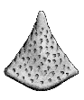
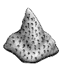
General Description
Male and female pelagic stingray teeth consist of a bilobate root and a cuspidate crown. The
teeth are small, ranging in width (mesio-distal) from 2 to 4 mm and depth (labio-lingual) from
1 to 2-1/2 mm. A moderately deep and wide nutrient groove is present between the two, lingually
directed, lobes. There is a single central pore which usually contains a single foramen. Lateral
foramina, on the interior surface of the lobes, have not been observed. The root lobes do not
extend beyond the lateral margins of the crown.
Anterior and lateral teeth of both sexes have cuspidate crowns, in contrast to the typical globular
design associated with most stingrays. The medial teeth have strong, lingually directed cusps,
which are similar to the "typical" high-cusped male stingray tooth -- such as those from
Dasyatis americana HILDEBRAND & SCHROEDER, 1928,
D. sayi (LESUER, 1817) and
D. sabina (LESUER, 1824).
Unlike the cited examples, the labial face does not have strong enameloid ridges or a
medial depression running the length of the cusp. In lateral teeth, the cusp is weaker and
more upright. In lieu of a cusp, the posterior teeth have a prominent, laterally elongated,
transverse crest which adjoins a strong transverse depression on the labial face. On the
lingual face, the transverse crest has longitudinal ridges. Medial and lateral teeth have a
weak depression on the labial face which is located at the juncture of the crown base and the cusp.
The labial face of all teeth have a pitted appearance which results from the depressions created
by numerous small meandering enameloid ridges. No teeth have a "cutting edge" as in many
shark or skate teeth. Rather, the acute angle of the transverse edge of the cusp and transverse
crest have a serrate edge created by the numerous enameloid ridges.
As a dasyatid, there is a unique feature represented in these teeth -- the development of a
second cusp. An incipient reflection of this characteristic can be seen in many teeth, usually
as a longitudinal depression in the lingual face of the crown. Eight percent of the teeth, originating
in medial and lateral positions, have a second cusp. These bicusped teeth appear sporadically
within the dentition, and are present in a file of teeth which are otherwise single-cusped. Unlike the
mobulids, the other family developing additional cusps, this bicuspidate condition in the pelagic
stingray is not associated with the mesio-distal elongation of the crown.
Sex and the isolated Pelagic stingray tooth
When studying the MBA shed tooth groups, certain information was available that would not be
available when examining isolated teeth of unknown origin. Of primary importance was the relative
sizes of male and female rays. The females were all larger, and examination by Greenwald &
Mollet confirmed that the teeth were also significantly larger in size. A second factor was the
availability of photographs of the dentitions themselves.
The anterior and most lateral teeth, as presented in this study, can be ascribed to a particular sex
with a strong degree of certainty. To a lesser extent and with less confidence, the posterior teeth
could be grouped by sex. This may not be the case when the origin of the teeth is unknown. If teeth
from a large male were intermixed with those of a small female, it is unlikely that any posterior, and
possibly lateral, teeth could be segregated. Anterior teeth from the first few files may show sexual
dimorphism.
I make the final comment from experience with the previously described dentition. Based on size,
and prior to extensive tooth study, that dentition was thought to be from a large male. The teeth
from that dentition were compared with the small (male) teeth collected from the aquarium. Although
smaller in size, these shed teeth all had counterparts in the dentition. When improved tooth collecting
techniques were developed, a new group of small teeth were identified -- they were clearly anterior
male teeth, but were not in the dentition. Once photographs of the MBA ray dentitions became
available, they were compared with the shed teeth and the studied dentition. It became readily
apparent that the dentition was from a small female rather than a large male. It was only the first few
files that had teeth sufficiently different to ascribe that difference to anything other than size.
Additional research may yield subtle morphological details which will allow teeth of unknown origin
to be confidently ascribed to a particular sex. However, at the time of this writing, relative size, a
very poor basis for identification, is the only characteristic available for most isolated teeth.
High-Cusped Teeth (Anterior)
Several distinct types of strongly cusped teeth can be found in the sample. Because observations
by MBA revealed that the female stingrays had much larger teeth than the males, it seemed relevant
to separate tooth-types by size.
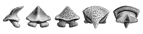
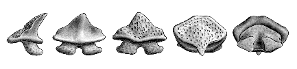
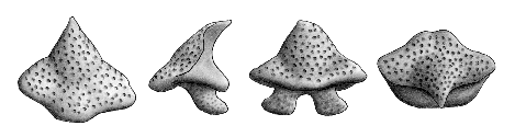
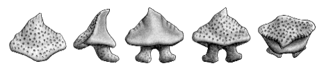
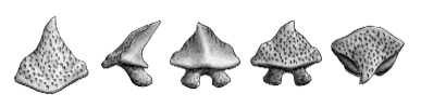
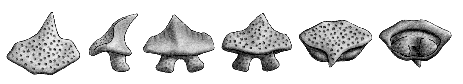
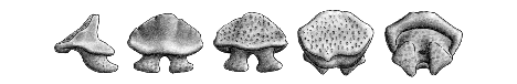
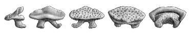
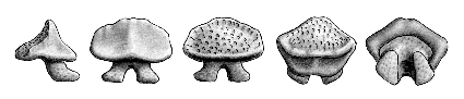
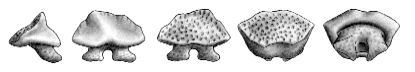
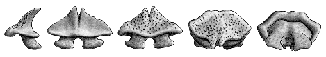
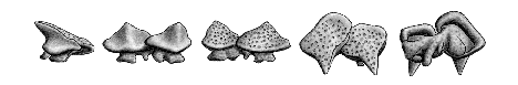
NOTE: All teeth were originally illustrated at 10X
and have maintained
a similar enlargement factor (to one another) when digitized. From
left to right, the aspects are: lateral, lingual, labial, occlusal and basal.
In certain cases, a cuspal view has been added to the left or one or more
aspects may have been omitted. |
- LARGE TEETH, probably shed by females.
 |
 |
| Cuspal view |
Specimen 54-C. hgt = 2.5, wid = 2.9,
dep = 2.5 mm |
- Type 54-C. These are one of the most common and identifiable of the pelagic stingray teeth.
In the March 14 sample, 12 of 136 teeth were segregated as belonging to this type and 10 of
those, were like the illustration. Even the sizes were consistent - WIDbc 3.0 (+/- 0.2) x
DEPbc 2.0 (+/- 0.1) mm. The triangular cuspal profile is characteristic, and the labial depression is
weak. There is a distinct labial flange, and the base of the crown, at the lateral angle, is high.
The cuspal angle is rounded and approximately 90 degrees.
 |
 |
| Cuspal view |
Specimen 54-B. hgt = 2.2, wid = 2.8.
dep = 2.0 mm |
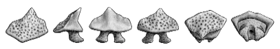
- Type 54-B. These teeth are similarly common but less easily identified -- their cuspal profile
and size tends to vary. Viewed labially, the cusp is often skewed to one side or the other but
the lateral profile remains stable. This tooth-type is characterized by its strong labial depression.
As with 54-C teeth, there is a distinct labial flange and high lateral angle. The March 14th sample had
examples (qty = 15) ranging from WIDbc 2.8 x DEPbc 2.0 to 3.9 x 2.5 mm. The cuspal angle
is rounded, slightly more than 90 degrees and located lower than noted on 54-C teeth.
A weak secondary cusp was observed on two specimens.
 |
| Specimen 58-A
hgt = 3.0, wid = 3.7, dep = 2.6 mm. |
- Type 58-A. The March 14th lot contained three teeth of this style. They are
generally similar to 54-B but the labial depression is very weak and the cuspal angle, higher.
These teeth were WIDbc 3.9 x DEPbc 2.8 and 3.4 x 2.0 mm in size.
 |
| Specimen 58-B
hgt = 2.8, wid = 4.0, dep = 2.6 mm. |
- Type 58-B. Similar to the preceding tooth-type, the March 14th lot contained
three teeth of this style. They are also similar to 54-B but the cuspal angle is very sharp and less
than 90 degrees. These teeth are WIDbc 4.0 x DEPbc 2.6, 3.9 x 2.3 and 2.9 x 2.2 mm in size.
One example is bicuspidate.
 |
| Specimen 60-C
hgt = 2.8, wid = 2.9, dep = 2.2 mm. |
- Type 60-C (was NYI-1). These teeth (two examples) appear to be fall between
54-B and 54-D. They have a strong labial depression, the cuspal angle is rounded and obtuse,
but the cusp remains lingually directed (but only slightly). In one example the cuspal angle is low
and in the other, high. These teeth are (WIDbc x DEPbc) 3.0 x 2.1 and 3.0 x 2.4 mm in size.
- SMALL TEETH, probably shed by males.
 |
| Specimen 58-D
hgt = 2.6, wid = 2.3, dep = 1.9 mm. |
- Type 58-D. Quite similar to the 54-C tooth described above, these
teeth are common (eleven in the March 14th sample) and generally triangular in shape.
However, the cusps are usually skewed to some extent. Other than one tooth measuring
2.5 x 1.8 (which is similar to this design but not exact), their size range is relatively consistent
(WIDbc x DEPbc) 2.3 (+/- 0.2) x 1.5 (+/- 0.1) mm. The labial flange is weak, cuspal angle obtuse
and labial depression moderate.
- Not Yet Illustrated - NYI-3. In this style tooth represented by three examples, the lower
labial face is broader and the cusp narrower -- a tear drop shape. The cusp is centered and the
labial depression moderate. The cuspal angle is rounded, low and obtuse. The labial flange
is strong and the crown base high at the lateral angle. The teeth measured
WIDbc 2.4 x DEPbc 1.5 (+/- 0.1) mm.
 |
| Specimen 58-C. hgt = 2.0, wid = 2.6, dep = 1.6 mm |
- Type 58-C. This tooth-type is a broader variation of NYI-3 but with a shorter cusp.
The labial flange is mild, labial depression weak and the rim rises at the lateral angle.
Ten teeth from the March 14th lot fell into this group. Between them there were differences
in the cuspal angle which coincided with size.
Six teeth have a cuspal angle which is rounded, obtuse and in a medium position. They
measured (WIDbc x DEPbc) 2.6 x 1.7, 2.6 x 1.6, 2.4 x 1.5, 2.3 x 1.6, 2.3 x 1.4 and 2.2 x 1.4 mm. The larger of
these teeth (illustrated) compares very well with upper teeth from files three and four
in the studied dentition. Three teeth had a less rounded cuspal angle which was slightly
higher and the cusp more lingually oriented. These teeth were 2.1 x 1.4 (+/- 0.1) in size.
The last example measured 2.3 x 1.4 but had a cusp more similar to NYI-3. Another example is bicusped.
 |
| Specimen 60-A. hgt = 1.9, wid = 2.1, dep = 1.6 mm |
- Type 60-A (was NYI-4). Viewed labially, these teeth
look similar to 58-C but with a lower cusp. Laterally however, the crown is upright with no pronounced
lingually directed cusp, somewhat similar to the 54-E design. This style tooth was represented by
five examples which measured WIDbc 1.9 (+/- 0.2) x DEPbc 1.2 (+/- 0.1) mm. The cuspal
angle is sharp and obtuse, with no apparent labial flange.
Low-Cusped Teeth (Lateral)
When viewed laterally, these teeth have a sharp apical surface, but there is no elongated cusp
directed lingually. Viewed labially or cuspally, the crown has an angular (pointed) apex which
provides a cusp-like appearance.
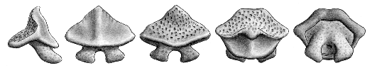 |
| Specimen 54-D. hgt = 2.8, wid = 3.5, dep = 2.3 mm |
- Type 54-D.
Viewed labially, these teeth have pointed crowns, but laterally, there is no obvious cusp -- the lateral profile
is somewhat triangular. Ten teeth from the March 14th lot fell into this grouping. There is a strong labial
depression which redirects the upper labial face and a rounded and obtuse lingual cusp angle which
creates an upright, cusp-like transverse crest. Sizes of these teeth are not uniform, ranging from
(WIDbc x DEPbc) 3.1 x 2.1 to 3.8 x 2.6 mm. (These large teeth were probably shed by females.)
Three of these teeth had two cusps. In one specimen (0314-122), one cusp was labially directed (54-D)
and the other, lingually (54-B).
 |
| Specimen 54-E. hgt = 2.0, wid = 2.8, dep = 2.1 mm |
- Type 54-E. This tooth-type compares well with teeth found in the lower lateral position of the
studied dentition. There is a transverse depression, labial flange and a moderately sharp cuspal
angle which slightly obtuse. Three teeth from the March 14th sample have been ascribe to this type, they
measured (WIDbc x DEPbc) 2.3 x 1.7 to 2.7 x 1.9 mm.
- NYI-6. Very similar to 54-E were five larger teeth: WIDbc 3.2 (+/- 0.1) x DEPbc 2.1 (+/- 0.2) mm.
Besides their larger size, the labial flange was often weaker, the cuspal angle sharper, and transverse
depression weaker. The cusp is low and lingually directed.
 |
| Specimen 55-B. hgt = 1.7, wid = 2.4, dep = 1.9 mm |
- Type 55-B. Originally lumped as a single tooth-type, there now appears to be two represented
by this design, one female (some upper teeth from the studied dentition look very much like these), and the
other possibly male. The sizes vary greatly. The illustrated example is (WIDbc x DEPbc) 2.5 x 2.1 mm
and the two similar teeth from the March 14th sample measure 3.4 x 2.2 and 3.0 x 2.1 mm. These teeth have
a strong labial flange, a cuspal angle in a medial position (rounded and obtuse) and high lateral angle.
- NYI-5. Very similar to the 55-B design, were three other teeth from the March sample. In these
teeth however, there is a sharp cuspal angle and the lingual face is more upright. (although the overall
crown height is lower). These teeth measured 3.0 x 2.2, 2.6 x 2.0 and 2.3 x 1.7 mm.
 |
| Specimen 60-B. hgt = 1.5, wid = 2.6, dep = 1.5 mm |
- Type 60-B (was NYI-10). Three teeth from the March 14th sample
are broad and low crowned and initially
might appear as posterior teeth. However, they were clearly cuspidate. Viewed laterally, the labially
face gently slopes in a lingual direction with a concavity in the area of the transverse depression. The
lingual face has an sharp and acute cuspal angle These teeth measured 2.6 x 1.4, 2.5 x 1.4 and
2.7 x 1.7 mm. (The later had two cusps.)
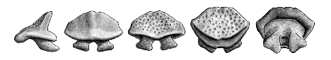
Non-Cuspidate Teeth (Posterior)
These teeth when viewed laterally have a relatively sharp apical angle, but when viewed from a
labial aspect, have little or no acute apical angle, but rather an elongated apical edge. Teeth
thought to be from distal positions of the dental band were originally placed into two groups, 54-A
and 55-D. This lumping was based on a morphological composite observable when viewed
laterally: 54-A teeth had a weakly concave labial face and an obtuse cuspal angle which gave
the lingual face a labial slope. 55-D teeth had a more strongly concave labial profile and a more
acute cuspal angle -- the upper lingual face is more upright. Since that time, 54-A teeth were found
to be less homogeneous in design than 55-D, and have been further refined.
In the studied dentition, the upper posterior teeth were of the broad 54-D design and the lower teeth
were similar to the 55-D tooth. Because of the variations in sizes within each group, it is likely that
male and female teeth are represented within these groups.
 |
| Specimen 54-A. hgt = 2.1, wid = 2.5, dep = 2.2 mm |
- Type 54-A. Only six specimens in the March sample correspond with the design
as illustrated above. Viewed lingually, the labially face is concave and the lingual slopes labially with a
rounded cuspal angle which is obtuse and in a median position. The lingual face above the cuspal
angle is labially directed. The crown has little or no labial flange and the transverse crest is broad
and straight. The WIDbc-DEPbc measurements range form 2.9 x 2.0 to 2.3 x 1.3 mm. A seventh tooth
has broadly separated roots and measures 2.9 x 1.5 mm - it may be pathological.
- NYI-7. Three teeth are very similar to the 54-A design but have a sharper and
less obtuse cuspal angle. This results in the upper lingual face being generally upright. These
teeth's basal crown measurements were 2.8 x 2.0 (+/- 0.1) mm.
 |
| Specimen 58-E
hgt = 1.9, wid = 2.7, dep = 2.0 mm. |
- Type 58-E. Six other teeth are similar to the 54-A design but have a less concave
labial face and a higher cuspal angle. These teeth fall into two distinct size (WIDbc-DEPbc) ranges:
(two teeth) 2.6 x 1.7 & 2.5 x 1.8 and (four teeth) 2.0 x 1.3 (+/- 0.1) mm.
- NYI-9. Two teeth are similar to the 54-A design but have a less concave labial face
and a low cuspal angle with a nearly upright, upper lingual face. These teeth measure
2.0 x 1.3 (+/- 0.1) mm.
 |
| Specimen 55-D. hgt = 1.5, wid = 2.1, dep = 1.6 mm |
- Type 55-D. The March 14th sample included twenty-two teeth with this design.
They represent some of the smaller teeth found, and likely originate in distal positions of the dental band.
The WIDbc-DEPbc measurements are quite erratic. If they originated from dental bands of significantly
different sizes (male and female), the graduation in sizes would be more logical. (Fourteen teeth ranged
from 2.1 x 1.3 to 2.3 x 1.8 mm, and eight measured 2.3 x 1.4 to 2.8 x 2.1 mm.) The study of a male dentition could
resolve this matter. In general, when viewed laterally, the labial face is concave, the transverse crest
upright and the lingual face has a sharp cuspal angle which is moderately obtuse. A labial flange is
generally lacking, and from a labial perspective, the crown appears neither cuspidate not pointed.
Bi-cuspidate Teeth
An unexpected morphological condition in this study was the presence of a second cusp in the teeth
of this species. In the preliminary sample from January, a single tooth was fully cuspidate
(illustrated in 55-A) but many (10%) displayed varying traces of this condition (incipient secondary
cusps, longitudinal depressions in the lingual face of the crown, etc.). In that January sample, this
condition was reflected in: 2/54-A, 1/54-B, 3/54-C, 1/54-D, 3/54-E, and 3/55-B teeth.
Since that time, a dentition was obtained which also reflected this characteristic. Because this
is no longer deemed a singular event (although both MBA rays and the studied dentition were
collected from a similar area and may reflect a characteristic of a localized population) only
teeth which have two cusps (and not the incipient condition) are now being counted. In the
0314 sample, 13 teeth had a second cusp (2/54-B, 4/54-D, 3/54-E, 58-B, 58-C?, NYI-08 & 60-B).
Looking at the distribution of bicusped teeth (using the allocation-to-tooth-group methodology
employed for the March 14 sample) within the dentition, nearly 25% of the lateral teeth display
this condition while only 6% of the anterior and 3% of the posterior teeth display this characteristic.
This could turn out to be a characteristic of lateral teeth only,
 |
| Specimen 55-A. hgt = 2.1, wid = 3.2, dep = 2.0 mm |
Pathological Teeth
Two examples were set aside in the first sampling as pathological teeth. One dual crowned tooth
(55-C, illustrated below) was clearly so, and the other had an inordinately broadened crown
and one disproportionately enlarged root lobe. The March sample produced four pathological
teeth. Specimen 0314-139 had three crowns, 0314-140, a laterally compressed tooth, a single root
lobe and 0314-138 was a bicusped tooth with pathological root lobes..
 |
| Specimen 55-C. hgt = 1.5, wid = 2.9, dep = 2.1 mm |
Condition
This aquarium sample provides shed teeth in superb condition.
Not only are they not subject to the depositional and weathering stress associated with
fossil teeth, but the rays themselves live the "Life of Riley" which includes their soft diet
of squid. Compared with the shed teeth in the described dentition, the MBA samples retain
many more features of the cusp.
Page revised July 31, 1997