|
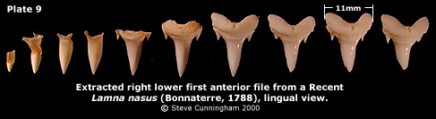
The C. taurus jaw used in this study had been cleaned of muscle and dermal tissue, then dried (see Pl. 1 in "Overview"). The youngest teeth of each file lack root structure in early stages of development (Pl. 9), resulting in their loss during cleaning due to weak attachment to the mucosa layer connecting them to the jaw.
Shark teeth do not arise from the jaw cartilage, they develop in collagen-rich mucosa within the anterior and lateroposterior hollows. If this connective tissue were to be completely removed, the crest of the jaw, where the functional teeth become erect, would show no evidence of having had teeth connected to it; teeth would be dangling from the bed of tissue. If the connective tissue were to disintegrate, teeth would be scattered. Materials used for tooth extraction include a turkey roasting pan, a food warming tray, some dental scrapers, and a large number of small plastic condiment cups such as those found in fast-food restaurants, labeled accordingly for each file of extracted teeth (68 cups were used here). Upon completion, the teeth were organized in a suitably sized Riker mount. The jaw was sawn into four pieces representing left and right, upper and lower halves and labeled accordingly using indelible ink. Each file was labeled by writing its position number on adjacent jaw cartilage. Teeth were extracted from one jaw section at a time. Anterior files were soaked for several minutes in water kept at 65 degrees Celsius (about 150 degrees Fahrenheit) in order to loosen weakly attached partially developed teeth found deep within the anterior hollow behind fully developed teeth, and removed with tweezers. Each tooth was placed in an appropriately labeled cup filled with hot water to further loosen recalcitrant tissue. Fully developed teeth, with well defined root structure, were soaked for a longer period of time and removed manually as they became loose enough to extract. Lateral and posterior teeth were extracted in like manner soaking only several files at a time. Upon completion of each jaw section, roots of fully developed teeth were scraped of all remaining tissue. The water in each cup was then replaced with 3% hydrogen peroxide solution and soaked for several hours to remove stains. The cups of teeth were emptied of solution and set aside to dry. Many of these characters may be observed on Pl. 6 (the horizontal dentition) and by using the Study Set. Overall tooth characters: Carcharias taurus has large narrow teeth bearing one sharp lateral denticle on each side of the base of the crowns. A distinct nutrient groove bisects the linqual root protuberance on all of the teeth. Roots are bilobed and crowns bear cutting edges which separate a strongly inflated lingual crown surface from a weakly inflated labial surface. The crowns are smooth except for occasional irregular, weak striations on lingual crown surfaces, especially near cutting edges. In increasingly distal files, tooth height lessens relative to width and lateral extension of the root lobes becomes equal to crown width resulting in tiny block-like teeth in distalmost files. Upper anterior teeth: Anterior teeth of the upper and lower jaw are long and slender for their width and have a very prominent lingual protuberance. In profile, upper anterior teeth are curved lingually with a strong labial recurvature. Lower anterior teeth are also curved lingually in profile, but very weakly recurved. See the Study Set to compare these characters of upper and lower anterior teeth. Profile view is an important factor sorting isolated fossil teeth to position. First upper anterior: Teeth of the first upper anterior
position have incomplete cutting edges which separate the strongly
inflated lingual surface of the crown from the weakly inflated
labial surface. The distal root lobe is equal or slightly longer
than the mesial. First upper anterior teeth are almost bilaterally
symmetrical when viewed lingually.
Intermediate teeth: The C. taurus jaw used for extraction had one file of intermediate teeth on each side of the upper jaw, originating from the bar of tissue separating the anterior and lateroposterior hollows. Careful examination of the jaw used for PL. 1 in "Overview" (a different jaw) reveals two intermediate files on the right side. Not shown, the left side of the same jaw bears three files of intermediate teeth. Intermediate teeth measure about one third the vertical height of third upper anteriors and have labio-lingually compressed root lobes. Root lobes are markedly rounded at the tips and the lingual surface of the lobes is flat. The narrow crown slants distally and in profile curves lingually, mildly recurved. The cutting edges are complete and the well-defined lateral denticles are triangular.
Upper lateroposterior teeth: Just as teeth originating in anterior hollows are termed "anteriors," those arising from the lateroposterior hollows are termed "laterals" and "posteriors." Upper lateral: Upper lateral teeth differ in shape from anterior teeth. Root lobe divergence is greater and remains constant throughout the successive files. Crowns are broader, flatter and shorter relative to root width in lingual view. Mesial root lobes are longer and distal root lobes are often broader, more rounded at the tip on the mesialmost files. As lateral files progress distally, root lobes become nearly bilaterally symmetrical. A single lateral denticle on each side of the base of the crown, while sharp, becomes noticeably triangular and flatter than those of anterior teeth. Cutting edges are complete on all lateral teeth. As lateral files progress distally, vertical crown height increases through the third file, then decreases as crowns become distally slanted. As lateral files progress to the posterior, inflation of the labial face of the enamel increases. Profile view reveals a less prominent lingual root protuberance than on anterior teeth. Crown profiles are flat with a mild labial recurvature. As lateral positions progress distally, labial inflation of the base of the crown increases. Upper posterior: Character gradation of lateroposterior positions is gradual throughout the entire palatoquadrate hollow, hindering a distinct division between lateral and posterior files. In this study, a vertical height/width ratio defines the division; posterior files have greater overall width than vertical height. In the Study Set, the eighth upper lateral also has a greater width than height, but retains its lateral designation due to a distinct blade and lateral denticles. In addition to a change in height/width ratio, the lateral denticles of posterior teeth loose their definition from the crown, becoming shoulder-like in appearance. Additionally, the vertical height of the crown equals that of the root, inflation of the basal labial enamel becomes very pronounced with distinctive wrinkles, and the lateral extension of root lobes disappears, becoming even with Crown width. Crown tips diminish giving the posterior teeth a block-like appearance.
Lower Anterior teeth: Lower anterior teeth are narrower, crowns more inflated and profiles more lingually curved than their upper counterparts. Mentioned in the upper anterior discussion, the lingually directed crowns have a very mild labial recurvature, a useful character distinguishing upper and lower anterior isolated fossil teeth. First lower anterior: Often termed "symphysial" or "parasymphysial" (see "Overview"), the first lower anterior teeth are markedly different from subsequent anteriors due to their size and asymmetry. The root lobes are compressed mesio-distally and convergent, pressed together except at the tips. The distal lobe is usually twice the length if the stunted mesial lobe. Vertical height is approximately one half that of the second lower anterior. A strong lingual protuberance overhangs the base of the lingual enamel. The cutting edges are incomplete. Second lower anterior: Teeth of this position resemble those of the first upper anterior position; root lobe divergence, general shape, a longer (or same length) distal root lobe, incomplete cutting edges and similar lateral denticles make them appear as large versions of first upper anteriors. These characters make sorting similar upper and lower isolated fossil teeth difficult. Mild labial recurvature of the crown tip and a deeper root lobe interspace distinguish second lower anteriors from first upper anteriors. Use the Study Set to compare these two positions. Roots bear a prominent lingual protuberance and the root lobes are compressed mesio-distally. Third lower anterior: Similar in appearance to second lower anteriors, teeth of this position are distinguishable by their noticeably longer and mesio-distally compressed mesial root lobe. The angle of root lobe divergence is greater than those of the second lower position. Fully developed third lower anteriors measure the greatest vertical height in the entire dentition. The cutting edges are incomplete. Fourth lower anterior: The angle of root lobe divergence is again greater than those of the previous positions. Use the horizontal dentition (Pl. 6 in "Dentition") or the Study Set to compare these angles as this relationship is very useful for sorting isolated fossil teeth. The longer mesial root lobe is somewhat compressed mesio-distally and the distal lobe is compressed labio-lingually. The crown profile curves lingually, as do teeth of the second and third positions, but differs by having a stronger labial recurvature. The cutting edges are incomplete.
Lower lateroposterior teeth: Height/width ratio of lower lateroposterior teeth distinguish them from anteriors, similar to upper lateroposteriors. However, flattening of the crown is not as pronounced and crown profiles show stronger lingual curvature than their upper counterparts. Use the Study Set to compare the differences of lingual curvature and recurvature for upper and lower lateroposterior teeth. Lower lateral: Angle of root lobe divergence is approximately equal to that of fourth lower anteriors throughout the lower lateral positions. Crowns are almost bilaterally symmetrical becoming distally slanted in distalmost files. The cutting edges are complete. Lower posterior: As with their upper counterparts, the height/width ratio, definition of lateral denticles and equal root/crown height distinguishes these teeth from lower laterals. Crowns loose tip definition in distalmost files and lateral root lobe extension becomes even with crown width. Basal labial inflation and wrinkling of the crown is very pronounced. Overview | Dentition | Study Set | Acknowledgements and References |