|
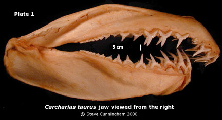
Arranging fossil shark teeth into artificial tooth sets requires an understanding of the relationships of teeth to each other and to their origin on the jaw. As fossil shark teeth are rarely associated with the jaw from which they arose, these relationships must be derived from the jaws and teeth of Recent sharks (see Agassiz,1835-1843; Leriche, 1902,1905; Applegate, 1965; Capetta, 1987; Ward, 1988; Kent, 1994; Kent & Powell, 1999; Siverson, 1999; and Cunningham, 2000). Prepared jaws (cleaned of tissue and dried, with teeth intact) of Recent sharks are used to study these relationships thereby limiting observation of tooth development and lingual surfaces of the root due to connective tissue and tooth proximity. While extracting one tooth (usually fully developed) from each file allows examination of lingual surfaces and positional characters, the entire file must be extracted in order to study developmental variation. Here, extracted files of teeth from a Recent specimen of Carcharias taurus (Pl. 1) are arranged in rows representing each position from both jaws (see Dentition menu). The jaws used in this study are from sandtiger sharks caught off the southern coast of Brazil, gender unknown. An interactive study set comprising one fully developed tooth from each file is a photo platform to show labial, profile, lingual views, and measurements (see "Study Set"). Teeth are displayed in life position, but when "clicked," the resulting photo shows crown pointing down for all positions facilitating comparison of upper and lower teeth.
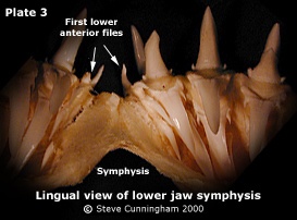
"Symphysial"
(proposed by Leriche, 1905), or more recently "parasymphysial"
(proposed by Cappetta, 1987), is often used for the small teeth
of the first lower anterior position in C. taurus. "First
lower anterior" is used here instead (Siverson, 1999). Applegate (1965, p. 6) cautions against abandoning established terms in order to avoid confusion, the benefit of which is lost if terms themselves are confusing. Cappetta (1987, p. 13) preserves the term "symphysial" for the first lower anteriors by adding the prefix "para" to distinguish them from unpaired teeth "astride" the symphysis, correctly pointing out that these teeth originate from either side of the lower symphysial bar. "Parasymphysial" has not been generally accepted as the term is inconsistent with terminology applied to files of larger teeth next to the symphysis in the upper jaw of C. taurus (i.e. first upper anterior). Confusion concerning
"symphysial" or "parasymphysial" results
from different perspectives: positional and functional. A positional
viewpoint allies the compressed, stunted teeth on either side
of C. taurus' lower symphysis with lower anterior files
as they share a common origin from within the lower anterior
hollows (Pl. 3, also see Siverson, 1999). A functional viewpoint
clearly distinguishes these teeth as different. Fossil evidence
suggests shark tooth function is in constant evolutionary transition.
Deriving tooth terminology from a functional point of view may
be less reliable considering, in evolutionary terms, any given
file of teeth can change function while maintaining the same
jaw position. Conversely,
the term "intermediate" accurately describes the small
teeth developing on the intermediate bar of tissue that separates
the anterior and lateroposterior hollows The term "Intermediate" is also (incorrectly) applied to the small files of anterior teeth harbored in the upper anterior hollows of Isurus oxyrinchus. Sharing their origin with first and second upper anterior teeth from the anterior hollows, referring to them as "third upper anteriors" eliminates inconsistent terminology regardless of their size (Siverson, 1999, text-fig. 3B). "File," "position" and "row" are sometimes interchanged when discussing tooth position (see "Roe vs Row" in the Reference page of elasmo.com). In this study, "file" represents functional and developing teeth (in line to replace the foremost teeth when shed) of any position while still on the jaw (Pl. 4). "Position" refers to successive files. "Row" is reserved for extracted teeth only, referring to a file of teeth arranged horizontally on a suitable surface for study. "Curvature" and "Recurvature" also have had ambiguous definitions in literature. Here, these terms apply to profile view. "Curvature" defines the direction the base of the crown is curving, usually lingually. "Recurvature" is reserved for occurrences where the tip of the crown curves in the opposite direction. "Slant" is used when teeth are no longer mesio-distally perpendicular to the base line of their root lobes (e.g. lateral teeth slant distally as files progress away from the midline of the jaw).
Orientation. Teeth are viewed lingually, crown tips pointing down. Often, crown tips are figured pointing up when shark teeth are depicted from directly above. However, while studying large numbers of fossil or extracted teeth, a viewpoint from directly above is impractical.Tooth characters such as compression of root lobes, shape/number of lateral denticles and striations are more easily recognized from a 45 degree line-of-sight lingual viewpoint when crown tips are pointing down. When extracted or fossil teeth are arranged in life position, lower teeth pointing up and upper teeth pointing down, differences in perspective make comparing characters of upper and lower teeth difficult. In "Dentition" the "horizontal dentition" arrangement eliminates viewpoint and perspective problems. Dentition | Study Set | Discussion | Acknowledgements and References |

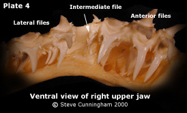 (Pl. 4, also Siverson, 1999, pp. 52-53,
text-fig. 3A).
(Pl. 4, also Siverson, 1999, pp. 52-53,
text-fig. 3A).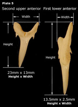 Measurements (Pl. 5) are expressed
height(mm) x width(mm) except as noted. Vertical height is represented
by a perpendicular line from the tangent of root lobe tips to
the crown tip. Width describes total width, usually the outside
width of root lobes. The mesio-distally compressed first lower
anterior teeth (often referred to as "symphysial" or
"parasymphysial") are measured by overall height and
width as root lobes are too irregular to determine a tangent.
Measurements (Pl. 5) are expressed
height(mm) x width(mm) except as noted. Vertical height is represented
by a perpendicular line from the tangent of root lobe tips to
the crown tip. Width describes total width, usually the outside
width of root lobes. The mesio-distally compressed first lower
anterior teeth (often referred to as "symphysial" or
"parasymphysial") are measured by overall height and
width as root lobes are too irregular to determine a tangent.