|
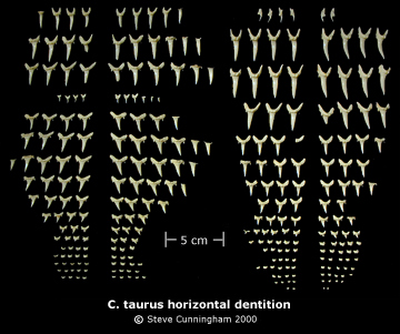
During study, keeping track of extracted or isolated fossil teeth is problematic due to numerous specimens and heterodonty in a single shark's dentition. Welton & Ferish (1993) defined three different types of tooth set: a "natural tooth set" where teeth and jaw more or less retain their original relationships (see Shimada, 1997), an "associated tooth set" involving teeth from a single individual but disassociated from the jaw (see Kent & Powell, 1999), and "artificial tooth set" for isolated teeth from various individuals of a single species (see Cunningham, 2000). In this study, "tooth set" is synonymous with "dentition." A partial or complete shark's dentition, where teeth are no longer attached to the jaw, is traditionally displayed in life position with the teeth of each file arranged vertically (in columns), anterior files in the middle and posterior files at either end. Upper jaw teeth are pointing down and, directly beneath, lower jaw teeth are pointing up. Arranged in this manner, positional comparisons are made difficult due to changes in perspective and excessive distances the eye has to travel. However, a horizontal arrangement of teeth from each file, crowns pointing in the same direction, eliminates perspective and distance problems by consolidating the dentition into a smaller area for study. The horizontal dentition (Pl. 6, also Cunningham, 2000) is proposed for practical construction and study of artificial tooth sets composed of numerous isolated fossil shark teeth. Sorting a single fossil species necessitates arranging teeth in unique rows representing similar shapes and characters. When Recent shark teeth are extracted and arranged similarly, positions of fossil rows become apparent by comparison.
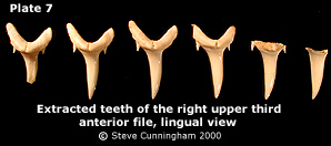
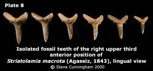
In the horizontal dentition, files extracted from the upper and lower jaw of C. taurus are arranged in horizontal rows, anterior teeth at the top, posterior teeth at the bottom. All crown tips are pointing down to enable easy comparison of upper and lower teeth. Upper teeth are arranged on the left half, lower jaw teeth on the right. Upper and lower jaw teeth are again separated into left and right halves. The centermost tooth of each half represents actual position, the foremost and most fully developed tooth in the file. Remaining teeth from each file are ranked according to development (see Pl. 7). Corresponding files of teeth on either side of the midline of the jaw, for any position, are arranged in a single horizontal row, as are corresponding upper and lower file positions, pointing in the same direction.
Pl. 6 disallows close-up examination of individual teeth. However, many positional characters are present that take advantage of the overall view: relative sizes, shapes, progressions, angles of root lobe divergence, damage and developmental stages. Additionally, when studying isolated fossil teeth, a horizontal dentition includes variational and ontogenetic characters. Overview | Discussion | Study Set | Acknowledgements and References |
||||||||||||||||||||